
Introduction
Clinical trials are designed to answer important questions about safety, efficacy, treatment response, and patient outcomes. To answer these questions reliably, research teams need accurate and measurable data. While lab values, clinical assessments, patient-reported outcomes, and safety reports are essential, imaging has become one of the most powerful sources of objective evidence. This is why medical imaging in clinical trials is now widely used across oncology, neurology, cardiology, orthopedics, respiratory studies, and many other therapeutic areas.
Imaging gives sponsors, CROs, investigators, radiologists, and clinical teams a visual way to evaluate what is happening inside the body. It can help identify disease status, confirm eligibility, measure progression, assess treatment response, and support endpoint evaluation. As a result, clinical trial imaging has become a critical part of modern research design and execution.
Why Imaging Is Important in Clinical Trials
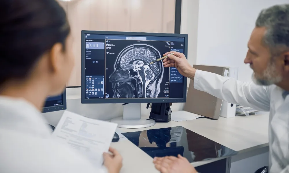
Medical imaging in clinical trials helps study teams collect visual and measurable information about a participant’s condition. Common imaging methods include CT, MRI, PET, ultrasound, X-ray, and other specialized imaging techniques.
In oncology trials, imaging is often used to measure tumor burden and evaluate whether a treatment is working. In neurology trials, MRI can help assess brain lesions, structural changes, or disease progression. In cardiology studies, imaging may support evaluation of heart function, blood flow, and vascular health. In orthopedic and musculoskeletal studies, imaging can help assess joint damage, bone healing, or tissue repair.
Because imaging provides objective evidence, it strengthens the overall quality of clinical trial data.
The Role of Clinical Trial Imaging in Patient Eligibility
Before a participant is enrolled, trial teams must confirm whether the patient meets protocol-defined eligibility criteria. Imaging can play an important role in this step.
For example, an oncology trial may require measurable disease at baseline. A neurology study may require MRI confirmation of disease severity. A cardiovascular trial may require imaging evidence of a specific structural or functional condition.
In these cases, clinical trial imaging helps ensure that enrolled participants match the intended study population. This reduces the risk of inappropriate enrollment and supports stronger trial validity.
Imaging and Treatment Response Assessment
One of the most important uses of medical imaging in clinical trials is treatment response assessment. Imaging allows researchers to compare scans taken before, during, and after treatment to understand whether the disease is improving, stable, or progressing.
In oncology, imaging criteria such as RECIST are often used to measure changes in tumor size. Radiologists compare baseline and follow-up images to classify response. In other therapeutic areas, imaging may measure inflammation, tissue changes, organ function, blood flow, or structural progression.
This makes imaging especially valuable when treatment effects cannot be fully understood through symptoms or lab values alone.
Why DICOM Matters in Imaging Data Management
As imaging data became more important in research, the need for standardization increased. This is where DICOM in clinical trials becomes essential. DICOM stands for Digital Imaging and Communications in Medicine. It is the standard format used to store, exchange, and manage medical imaging data.
DICOM medical imaging includes both the image and important metadata. This metadata may include scan date, modality, image orientation, scanner details, acquisition parameters, study identifiers, and patient-related information.
In clinical trials, imaging data may come from many sites, hospitals, scanners, and countries. Without a standard such as DICOM, it would be difficult to organize, compare, and review imaging data consistently.
DICOM Medical Imaging and Data Quality
DICOM medical imaging supports better data quality by preserving important technical and study-related information. This helps imaging teams confirm whether scans were captured according to protocol requirements.
For example, if a trial requires a specific MRI sequence or CT acquisition protocol, DICOM metadata can help reviewers verify whether the submitted image meets the requirement. If metadata is missing or incorrect, the image may be difficult to review or compare.
Using DICOM in clinical trials also supports traceability. It helps teams connect images to the correct subject, visit, timepoint, and study while supporting secure review workflows.
Common Challenges in Clinical Trial Imaging
Despite its value, clinical trial imaging can be difficult to manage. Imaging files are large, and trials may require repeated scans across several visits. Secure storage, transfer, upload, and review workflows must be carefully managed.
Another challenge is site variation. Different sites may use different scanners, acquisition settings, and imaging workflows. If sites do not follow the imaging protocol consistently, image quality and comparability may be affected.
De-identification is another critical requirement. DICOM files may contain patient identifiers in metadata fields, so images must be anonymized before central review or analysis. At the same time, essential trial information must be preserved so that images remain traceable within the study.
The Importance of Central Imaging Review
Many trials use central imaging review to improve consistency and reduce bias. In this model, images collected from trial sites are reviewed by independent radiologists or imaging experts using standardized criteria.
Central review is especially important when imaging contributes to primary or secondary endpoints. It helps ensure that images are assessed consistently, regardless of where they were captured.
A strong central review process depends on complete, high-quality, properly de-identified DICOM data. Without good imaging data management, review timelines can be delayed and endpoint reliability may be affected.
How AI Is Supporting Imaging Workflows
AI is increasingly being explored in imaging workflows. It can help with image quality checks, lesion detection, segmentation, measurement support, anonymization review, and imaging biomarker analysis.
AI can also help manage large imaging datasets by identifying images that may need closer review or by supporting more consistent measurements. However, AI depends on structured and high-quality data. This makes DICOM medical imaging and strong imaging governance even more important.
AI should support radiologists and imaging experts, not replace them. Human expertise remains essential for interpretation, validation, and clinical decision-making.
Conclusion
This bloggingwebs article must have given you a clear understanding of the topic. Medical imaging in clinical trials plays a major role in patient selection, disease monitoring, treatment response assessment, and endpoint evaluation. It provides visual and measurable evidence that can strengthen the reliability of trial results.
DICOM in clinical trials provides the standard structure needed to manage imaging data across sites, systems, and reviewers. With strong DICOM medical imaging workflows, sponsors and CROs can improve traceability, image quality, and review consistency.
As clinical research becomes more data-driven, strong clinical trial imaging processes will become even more important for generating reliable evidence and supporting better trial decisions.