In regenerative tissue engineering, ischemic wound healing, and vascular biology, restoring functional blood flow is the fundamental rate-limiting step. Without a functional microvascular network, newly engineered tissues quickly suffer from localized hypoxia, nutrient deprivation, and metabolic waste buildup.
Angiogenesis—the physiological process through which new capillary blood vessels sprout from pre-existing vasculature—requires precise chemical coordination. Endothelial cells must degrade the surrounding basement membrane, migrate into the extracellular matrix, proliferate, and assemble into hollow capillary tubes.
While large recombinant proteins like Vascular Endothelial Growth Factor ($VEGF$) can trigger vessel sprouting, their short half-lives and potential to induce uncontrolled vascular permeability present operational challenges in long-term models.
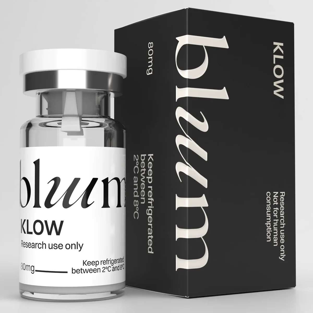
Consequently, researchers are actively evaluating synthetic biomimetic sequences designed to target specific endothelial cell receptors. Studying how the synthetic klow peptide derivative interacts with human umbilical vein endothelial cells (HUVECs) provides essential insights into targeted microvascular formation and tubulogenesis.
1. Cellular Mechanics of Capillary Tube Sprouting
Building a functional capillary network requires a tightly regulated sequence of cellular events. When endothelial cells receive an pro-angiogenic signal, they undergo distinct morphological changes:
- Endothelial Tip Cell Selection: A single endothelial cell responds to local signal gradients, assuming a motile “tip cell” phenotype characterized by long filopodia that sense the extracellular environment.
- Extracellular Matrix Degeneration: Tip cells secrete matrix metalloproteinases (primarily MMP-2 and MMP-9) to locally degrade the basement membrane, creating a pathway for sprout elongation.
- Stalk Cell Proliferation and Lumen Formation: Trailing “stalk cells” proliferate rapidly behind the tip cell, forming a continuous cord. Intracellular vacuoles then coalesce across adjacent cells, generating a hollow vascular lumen.
- Pericyte Recruitment: Maturing vessels secrete PDGF-$\beta$, recruiting pericytes and smooth muscle cells to stabilize the new vessel wall.
2. Intracellular Signaling Networks in Peptide-Driven Tubulogenesis
Synthetic bio-active sequences stimulate tubulogenesis by engaging cell-surface integrins and endothelial growth factor receptor complexes.
The intracellular signaling cascade follows a structured biological sequence:
1. Endothelial Cell Surface Engagement:Receptor Binding.
The bio-active peptide binds to cell-surface integrin complexes ($\alpha_v\beta_3$) and co-receptors, inducing conformational changes that autophosphorylate VEGFR2 tyrosine residues.
2. eNOS Phosphorylation via Akt:Enzymatic Activation.
Downstream signaling activates the PI3K/Akt pathway, phosphorylating endothelial Nitric Oxide Synthase (eNOS) at Ser1177 to increase local nitric oxide ($NO$) production.
3. FAK Activation and Filopodia Extension:Cytoskeletal Reorganization.
Nitric oxide and focal adhesion kinase (FAK) signaling trigger F-actin filament reorganization, driving filopodia formation and endothelial cell migration into the matrix core.
4. Lumen Coalescence and Branching:Tube Assembly.
Migrating endothelial cells align end-to-end, forming intercellular junctions and hollow lumens that join into interconnected vascular networks within 12 to 18 hours.
3. Measuring Angiogenic Metrics in Endothelial Models
Quantifying angiogenic efficacy in vitro requires tracking key morphological parameters in HUVEC tube formation assays on basement membrane matrices:
Quantitative assays demonstrate that applying the klow peptide derivative to endothelial cultures accelerates capillary loop closure and increases branching density, establishing a robust microvascular network in experimental models.
4. Analytical Quality Controls for Vascular Assay Reagents
Vascular assays are sensitive to minor chemical variations. Residual synthesis contaminants—such as trifluoroacetic acid (TFA) salts or truncated deletion sequences—can cause endothelial cell detachment, vessel degradation, or uncharacterized inflammatory responses.
To obtain clean, reproducible data, research protocols require high-purity reagents verified by tandem mass spectrometry (MS/MS) and analytical high-performance liquid chromatography (RP-HPLC). Utilizing certified, high-purity klow peptide materials ensures that measured increases in vessel branching reflect genuine pro-angiogenic activity rather than experimental noise.
5. Advancing Microvascular Research in Tissue Engineering
Establishing functional vascular networks is essential for advancing tissue engineering and regenerative therapies. Biomimetic signaling sequences provide a targeted, controllable method for stimulating endothelial tube formation and capillary sprouting.
Continued investigation into the cellular mechanisms of the klow peptide derivative deepens our understanding of receptor-mediated tubulogenesis. Grounding these studies in rigorous analytical standards ensures that synthetic reagents deliver clean, reproducible, and publication-ready results across every stage of vascular discovery.